Anatomy Of Chest X Ray / Normal Chest X Ray Anatomy Tutorial Kenhub - It is almost always the first imaging study ordered to evaluate for pathologies of the thorax, although further diagnostic imaging, laboratory tests.
byAdmin•
0
Anatomy Of Chest X Ray / Normal Chest X Ray Anatomy Tutorial Kenhub - It is almost always the first imaging study ordered to evaluate for pathologies of the thorax, although further diagnostic imaging, laboratory tests.. • the straight back syndrome or pectus. The interpretation of a chest film requires the understanding of basic principles. Air spaces normally seen in. Posted by radiologypics ⋅ march 17, 2013 ⋅ leave a comment. Published 2011 by blackwell publishing ltd.
Gillian lieberman forthe harvard 62. Conclusion of living anatomy of the chest congratulations! Chest radiographs are the most common film taken in medicine. • the straight back syndrome or pectus. Evaluation of a chest radiograph may appear to be simple, but is in fact a complex task requiring careful observation, sound understanding of chest anatomy, and knowledge of the principles of physiology and pathology.
The Chest X Ray Ppt Video Online Download from slideplayer.com • the straight back syndrome or pectus. Many clinical conditions can be evaluated by this simple radiology test. You have completed this module. L the portion of the left lung that corresponds anatomically to the right middle lobe is incorporated into the left upper lobe. Because some conditions of the chest. The interpretation of a chest film requires the understanding of basic principles. Air spaces normally seen in. Posted by radiologypics ⋅ march 17, 2013 ⋅ leave a comment.
In this article we will focus on:
It is almost always the first imaging study ordered to evaluate for pathologies of the thorax, although further diagnostic imaging, laboratory tests. Because some conditions of the chest. It first appears too complicated to read the chest xrays because we barely know what. In fact every radiologist and pulmonary physician should be an expert in chest film reading. You have completed this module. It is used to evaluate the lungs, heart and chest what are the limitations of chest radiography? Labeled chest radiographs teaching radiologic anatomy with a level of detail appropriate for medical students. Air spaces normally seen in. Gillian lieberman forthe harvard 62. Conclusion of living anatomy of the chest congratulations! Evaluation of a chest radiograph may appear to be simple, but is in fact a complex task requiring careful observation, sound understanding of chest anatomy, and knowledge of the principles of physiology and pathology. Living anatomy of the chest for 1st year medical students original version compiled by dr. Doctors use them to diagnose problems.
Abcde aproach the anatomy of the heart can appear artificially larger due to this image orientation. The interpretation of a chest film requires the understanding of basic principles. Look for lung and pleural pathology. Gillian lieberman forthe harvard 62. Because some conditions of the chest.
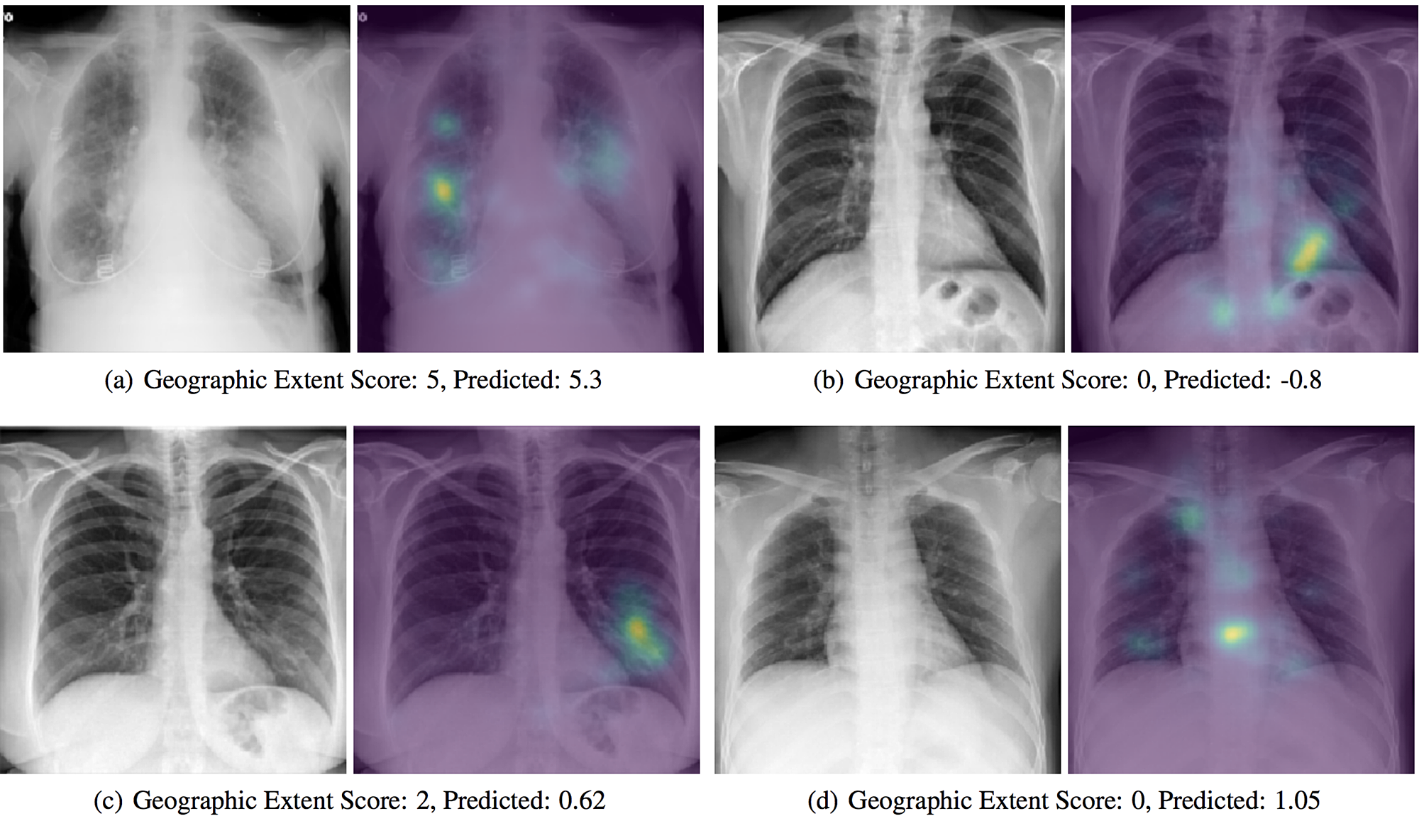
Cureus Predicting Covid 19 Pneumonia Severity On Chest X Ray With Deep Learning from assets.cureus.com This imaging method can also check how a patient is responding to specific treatments. Chest radiographs are the most common film taken in medicine. Because some conditions of the chest. The interpretation of a chest film requires the understanding of basic principles. In fact every radiologst should be an expert in chest film reading. Evaluation of a chest radiograph may appear to be simple, but is in fact a complex task requiring careful observation, sound understanding of chest anatomy, and knowledge of the principles of physiology and pathology. Each of these anatomical structures should be viewed using a systematic approach. It is used to evaluate the lungs, heart and chest what are the limitations of chest radiography?
Labeled chest radiographs teaching radiologic anatomy with a level of detail appropriate for medical students.
Medzcool On Twitter Can You Identify And Name All These Structures On A Chest X Ray Learn More About The Anatomy Of A Chest X Ray Here Https T Co 9ucnjtxkex Radiology Anatomy Medstudent Medicalstudent Usmle Nursing from pbs.twimg.com Because some conditions of the chest. Common symptoms that can be diagnosed using chest. Evaluation of a chest radiograph may appear to be simple, but is in fact a complex task requiring careful observation, sound understanding of chest anatomy, and knowledge of the principles of physiology and pathology. Labeled chest radiographs teaching radiologic anatomy with a level of detail appropriate for medical students. Doctors use them to diagnose problems. Living anatomy of the chest for 1st year medical students original version compiled by dr. However, finding problems that are often a/w arrhythmias, such as cardiac enlargement and lung disease, should alter one to the possibility of arrhythmias. In fact every radiologist and pulmonary physician should be an expert in chest film reading.
Look for lung and pleural pathology.
Many clinical conditions can be evaluated by this simple radiology test. Major structures are shown in fig. Evaluation of a chest radiograph may appear to be simple, but is in fact a complex task requiring careful observation, sound understanding of chest anatomy, and knowledge of the principles of physiology and pathology. Chest radiographs are the most common film taken in medicine. In fact every radiologst should be an expert in chest film reading. You have completed this module. Air spaces normally seen in. A method for examining a chest. Posted by radiologypics ⋅ march 17, 2013 ⋅ leave a comment. It is almost always the first imaging study ordered to evaluate for pathologies of the thorax, although further diagnostic imaging, laboratory tests. Is there any inhaled foreign body? L the portion of the left lung that corresponds anatomically to the right middle lobe is incorporated into the left upper lobe. Because some conditions of the chest.
In fact every radiologst should be an expert in chest film reading anatomy of chest. The interpretation of a chest film requires the understanding of basic principles.